Cytochrome c oxidase (CcO) — also known as Complex IV of the mitochondrial electron transport chain — is the primary photoacceptor in red and near-infrared light therapy. When photons in the 600–900 nm range reach the mitochondria, CcO absorbs them, triggering three key responses: increased ATP (cellular energy) production, release of nitric oxide (a vasodilator that improves blood flow), and modulation of reactive oxygen species that activates protective signaling pathways. This mechanism, first identified by Karu et al. (2005), is what makes photobiomodulation work at the molecular level.
Introduction
If photobiomodulation were a car, cytochrome c oxidase would be the ignition switch. Without this specific molecular target absorbing light energy, the cascade of beneficial cellular effects — from collagen production to pain relief — simply wouldn’t occur.
The identification of CcO as the primary chromophore for PBM was a watershed moment. Through the work of Tiina Karu and colleagues, published in a landmark 2005 study (Karu et al., 2005), researchers finally had a molecular explanation for what Endre Mester had observed nearly four decades earlier: that low-intensity red light could stimulate biological processes in living tissue.
Yet many device manufacturers and consumers remain unaware of why specific wavelengths work — and why others don’t. This article explains the molecular mechanism that makes red light therapy possible, from photon absorption to downstream biological effects.
Understanding this mechanism matters for three audiences:
- B2B clients and OEM partners: CcO science directly informs device wavelength selection, irradiance calibration, and evidence-based marketing claims [[4]][doc_4]
- Healthcare professionals: it provides the mechanistic framework for understanding clinical outcomes
- Informed consumers: it offers confidence in the science behind the products they use
At WakeLife Beauty, our device engineering is guided by this fundamental science — ensuring that wavelength selection and power output parameters are optimized for effective CcO activation.
What is Cytochrome c Oxidase?
The Final Enzyme in Cellular Respiration
Cytochrome c oxidase is the fourth and final enzyme complex of the electron transport chain (ETC), located in the inner mitochondrial membrane. It performs a critical function: catalyzing the final step of cellular respiration by transferring electrons to molecular oxygen while pumping protons across the membrane. This proton gradient drives ATP synthase, producing the vast majority of the cell’s ATP through oxidative phosphorylation (Chung et al., 2012).
What makes CcO uniquely relevant to photobiomodulation is that it contains chromophores — light-absorbing molecular components — that happen to absorb wavelengths in the red and near-infrared spectrum.
Structure and Chromophores
CcO is a large transmembrane protein complex containing multiple metal centers that serve as chromophores:
| Chromophore | Location | Peak Absorption Region | Function in ETC |
|---|---|---|---|
| Heme a | Subunit I | ~600 nm | Electron transfer |
| Heme a3 | Subunit I | ~600–605 nm | Oxygen binding site |
| CuA center | Subunit II | ~820–830 nm | Initial electron acceptor |
| CuB center | Subunit I | ~600–605 nm | Coupled to heme a3 |
Source: Chromophore identification and absorption characteristics established by Karu et al. (2005).
These metal centers — particularly the copper and heme groups — are the reason red and near-infrared light can interact with mitochondria. Without them, light at these wavelengths would simply pass through the cell with no biological effect.
How Does CcO Absorb Light? The Photochemical Mechanism
When red or near-infrared light reaches the mitochondria, a four-step sequence occurs:
Step 1 — Photon Absorption
Photons in the 600–900 nm range penetrate tissue and reach mitochondrial membranes. CcO’s chromophores absorb specific wavelengths based on their electronic structure:
- Heme centers absorb primarily in the red range (~600–660 nm)
- CuA centers absorb primarily in the near-infrared range (~810–850 nm)
This dual absorption profile explains why devices using both red and NIR wavelengths provide more comprehensive CcO activation (Karu et al., 2005).
Step 2 — Nitric Oxide Dissociation
Under normal cellular conditions, nitric oxide (NO) binds to CcO at the oxygen binding site (heme a3/CuB), competitively inhibiting the enzyme’s ability to reduce oxygen. This is a natural regulatory mechanism — but it also means CcO often operates below its maximum capacity.
When photons are absorbed by CcO, the energy causes NO to dissociate from the enzyme. This “unclogging” has two important effects:
- CcO is freed to operate at full efficiency — electron transport accelerates
- The released NO becomes a signaling molecule — causing vasodilation and improved blood flow
Poyton & Ball (2011) proposed a detailed mechanistic model explaining how this light-induced NO release from CcO leads to both mitochondrial and transcriptional effects — connecting a single photochemical event to diverse downstream outcomes.
Step 3 — Enhanced Electron Transport and ATP Synthesis
With NO removed, the electron transport chain operates more efficiently:
- Electron transport rate increases
- Proton pumping across the mitochondrial membrane accelerates
- The electrochemical gradient strengthens
- ATP synthase produces more ATP
Mochizuki-Oda et al. (2002) directly measured increased ATP production in mitochondria exposed to near-infrared laser irradiation. Chung et al. (2012) reviewed the broader evidence across multiple experimental models confirming this effect.
A note on ATP increase claims: You may see some sources cite specific percentage increases in ATP (e.g., “150–200%”). The reality is more nuanced: the magnitude of ATP increase varies significantly depending on cell type, baseline metabolic state, wavelength, and dosage. The evidence consistently shows that cells in a stressed or compromised state tend to show the largest ATP response to PBM, while already-healthy cells show more modest changes (Chung et al., 2012). This is consistent with PBM being primarily restorative rather than supraphysiological — it helps struggling cells recover, rather than pushing healthy cells beyond their normal output.
Step 4 — Downstream Signaling Cascades
The changes in ATP, reactive oxygen species (ROS), and NO trigger multiple signaling pathways over hours to days:
| Pathway | Full Name | Primary Effect in PBM |
|---|---|---|
| NF-κB | Nuclear factor kappa-B | Anti-inflammatory response (Hamblin, 2017) |
| MAPK/ERK | Mitogen-activated protein kinase | Cell proliferation, tissue repair |
| PI3K/Akt | Phosphoinositide 3-kinase | Cell survival, cytoprotection |
| Nrf2 | Nuclear factor erythroid 2-related factor 2 | Antioxidant defense upregulation |
Pathways reviewed in Chung et al. (2012) and Hamblin (2017).
These pathways are covered in detail in our dedicated article: → Topic 04: Downstream Effects of PBM — ATP, Inflammation & Antioxidant Defense [[1]][doc_1]
Wavelength Specificity: Why Not All Light Works
CcO’s chromophores have specific absorption peaks. This means wavelength selection is not arbitrary — it’s dictated by molecular physics.
| Wavelength Range | Primary CcO Target | Tissue Penetration | Best For |
|---|---|---|---|
| 630–660 nm (Red Light) |
Heme a, Heme a3 | ~1–3 mm(epidermis, dermis) | Skin rejuvenation, surface wounds, dermatological conditions |
| 810–850 nm (Near-Infrared) |
CuA center | ~3–10 mm(muscle, tendon, nerve) | Pain, joints, muscle recovery, neurological applications |
Absorption peaks: Karu et al. (2005). Penetration depth estimates: Chung et al. (2012) reviewing tissue optics literature.
What About Other Wavelengths?
Not all light interacts with CcO:
- UV light (< 400 nm): Does not activate CcO. Damages DNA through a completely different mechanism. Not therapeutic — it’s harmful.
- Blue light (400–480 nm): Targets different chromophores (primarily flavins and porphyrins). Used in acne treatment, but through a different biological pathway. Not a CcO activator [[2]][doc_2].
- Green/yellow light (500–600 nm): Minimal CcO absorption. Very limited PBM evidence.
- Above 900 nm: Water absorption in tissue increases significantly in this range, reducing the proportion of photons that actually reach CcO. While CcO’s copper centers have some theoretical absorption at these wavelengths, the practical efficiency drops considerably. The clinical evidence base concentrates overwhelmingly on the 600–850 nm range.
This is why the dual-wavelength approach (660 nm + 830–850 nm) has become the standard in well-designed PBM devices: it targets both major chromophore groups within CcO while staying in the wavelength range where tissue penetration is most efficient.
→ For comprehensive wavelength analysis: Topic 06: Wavelength Selection & Tissue Penetration Depth in PBM Devices [[1]][doc_1]
Clinical Evidence for CcO as the Primary Photoacceptor
The identification of CcO as PBM’s molecular target rests on multiple converging lines of evidence:
Karu et al. (2005) — The Definitive Identification
Tiina Karu and colleagues demonstrated that the action spectrum of PBM (which wavelengths produce biological effects) closely matches the absorption spectrum of oxidized CcO. This spectral matching was the critical evidence linking PBM to a specific molecular target — and it explained why certain wavelengths work while others don’t.
Poyton & Ball (2011) — The NO–CcO Mechanistic Model
Robert Poyton and Katelyn Ball proposed the most detailed mechanistic model explaining how light-induced NO release from CcO leads to both immediate mitochondrial effects (ATP increase) and longer-term transcriptional changes (gene expression). Their model explains how a single photochemical event — NO dissociation — can produce diverse downstream outcomes across different tissues.
Mochizuki-Oda et al. (2002) — Functional Evidence: ATP and Blood Flow
This study directly measured increased ATP production in mitochondria and improved cerebral blood flow following near-infrared irradiation — providing functional (not just spectroscopic) evidence that CcO activation translates to measurable physiological outcomes.
Wang et al. (2017) — CcO Mechanism in Brain Applications
A comprehensive study demonstrating that near-infrared light (810 nm) can modulate brain function through transcranial delivery, with CcO activation as the proposed mechanism. This work extended the CcO model from skin and muscle into neurological applications.
Chung et al. (2012) — The Definitive PBM Review
The most-cited review in PBM literature (2,000+ citations) synthesizes the complete evidence for CcO as the primary chromophore and maps the full mechanistic chain from photon absorption to clinical effects. This paper remains the essential reference for anyone seeking to understand PBM’s molecular basis.
Implications for Device Design
Understanding CcO science has direct, practical implications for how PBM devices should be designed, evaluated, and selected [[1]][doc_1].
Wavelength Selection
Based on CcO absorption spectra:
- 660 nm (red): Targets heme centers. Optimal for skin-depth applications.
- 830–850 nm (NIR): Targets CuA center. Optimal for deeper tissue applications.
- Dual-wavelength (660 + 850 nm): Comprehensive CcO activation across tissue depths. This has become the evidence-based standard.
Irradiance and Dosimetry
CcO activation follows a biphasic dose response — a critical concept covered in Topic 03: Biphasic Dose Response [[4]][doc_4]:
| Parameter | Effective Range | Rationale |
|---|---|---|
| Irradiance | 30–100 mW/cm² | Sufficient photon flux to activate CcO without thermal effects |
| Energy Density | 3–10 J/cm² (superficial); higher for deep targets | Balance between activation and saturation |
| Treatment Duration | 10–20 minutes typical | Dependent on irradiance and target dose |
Dosimetry parameters based on ranges reviewed in Chung et al. (2012) and Hamblin (2017).
Key principle: More light is not always better. Exceeding optimal doses can shift cellular responses from stimulatory to inhibitory (Huang et al., 2009). This is why device specifications — not just “having red LEDs” — determine clinical outcomes.
→ Full dosimetry guide: Topic 07: Irradiance, Energy Density & Dosimetry [[1]][doc_1]
How This Science Informs Our Device Engineering
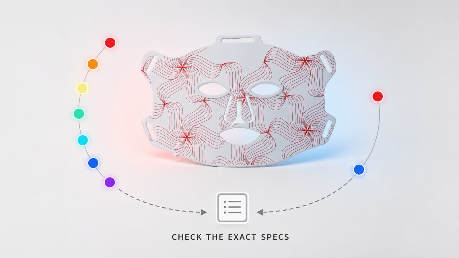
At WakeLife Beauty, CcO absorption science directly guides product development. Our LED face masks use 660 nm + 850 nm dual wavelengths — not because it’s a marketing trend, but because these wavelengths correspond to the peak absorption of CcO’s heme centers and CuA centers respectively, providing activation across tissue depths [[3]][doc_3].
Irradiance is calibrated within the effective range identified in the research cited throughout this article, balancing CcO activation against the biphasic dose response. This is the difference between a science-informed device and a generic LED product.
→ Explore our product range: LED Face Masks | LED Therapy Panels → For OEM/ODM partnerships: Custom Device Development [[3]][doc_3]
Common Misconceptions
“Any red light will work”
Reality: Only wavelengths in the 600–900 nm range effectively activate CcO’s chromophores (Karu et al., 2005). A red decorative light, a heat lamp, or an infrared sauna operate at wavelengths or power levels that do not match CcO’s absorption spectrum. Wavelength specificity is dictated by molecular physics, not marketing preference.
“Brighter is always better”
Reality: CcO activation follows a biphasic dose response. Beyond the optimal irradiance range, additional photons can actually inhibit cellular function rather than enhance it (Huang et al., 2009). This is why reputable device manufacturers specify irradiance ranges rather than just advertising maximum power.
“All red light therapy devices are the same”
Reality: Wavelength accuracy, spectral bandwidth, irradiance uniformity, and thermal management vary enormously between devices. A device emitting at 620 nm instead of 660 nm will have different CcO absorption efficiency. A device with poor thermal management may shift wavelength output as it heats up. These engineering details directly affect whether CcO is being optimally activated — and they’re invisible to the consumer without proper documentation.
FAQ
What is cytochrome c oxidase and why does it matter for red light therapy?
Cytochrome c oxidase (CcO) is the enzyme in your mitochondria that absorbs red and near-infrared light. It’s the molecular reason photobiomodulation works. When CcO absorbs photons in the 600–900 nm range, it produces more cellular energy (ATP) and releases nitric oxide — triggering the healing, anti-inflammatory, and regenerative effects associated with red light therapy (Karu et al., 2005; Chung et al., 2012).
Which wavelengths best activate CcO?
CcO has two primary absorption regions: approximately 660 nm (red light, targeting heme centers) and 830–850 nm (near-infrared, targeting copper centers). Devices using both wavelengths provide the most comprehensive CcO activation (Karu et al., 2005).
How quickly does CcO respond to light?
CcO absorbs photons and releases NO within seconds. ATP increases are measurable within minutes. However, the full downstream effects — changes in gene expression, collagen synthesis, inflammatory markers — develop over hours to days with consistent treatment.
Can CcO be over-activated?
Yes. CcO follows a biphasic dose response: moderate doses stimulate, excessive doses inhibit (Huang et al., 2009). This is why device dosimetry (irradiance × time = energy density) matters — and why “more powerful” doesn’t automatically mean “more effective.” See Topic 03: Biphasic Dose Response for details.
Does CcO explain all of PBM’s effects?
CcO is the primary mechanism and explains the majority of PBM’s documented effects. Other chromophores — including flavins, opsins, and water molecules at specific wavelengths — may contribute to certain effects. But the scientific consensus, established by Karu (2005) and reinforced by subsequent research, is that CcO is the dominant photoacceptor for red and NIR wavelengths (Chung et al., 2012).
How is red light therapy different from UV light or tanning?
Completely different. UV light (below 400 nm) damages DNA and does not interact with CcO. Red and NIR light (600–900 nm) is absorbed by CcO and triggers protective, restorative cellular responses. Their biological effects are essentially opposite.
Conclusion
Cytochrome c oxidase is the molecular gateway through which photobiomodulation exerts its effects. The mechanism — photon absorption → NO dissociation → enhanced electron transport → increased ATP → downstream signaling — is well-established through decades of research from Karu, Poyton, Hamblin, Chung, and others.
For anyone evaluating, designing, or using PBM devices, CcO science answers the most fundamental question: why does this work?
And from that understanding flow practical decisions:
- Wavelength selection: 660 nm + 830–850 nm targets CcO’s two major chromophore groups
- Irradiance parameters: 30–100 mW/cm² provides sufficient photon flux for CcO activation
- Dose management: The biphasic response means proper dosimetry is non-negotiable
- Quality differentiation: Wavelength accuracy and spectral purity directly affect CcO activation efficiency
As PBM research continues — particularly in neurology, immunology, and metabolic applications — our understanding of CcO’s role will deepen. But the core mechanism is established science, not speculation. And it’s this science that should guide every device that claims to deliver red light therapy [[4]][doc_4].
Continue reading:
- → Topic 01: Photobiomodulation — Definition, History & How It Works
- → Topic 03: Biphasic Dose Response — Why More Light Is Not Always Better
- → Topic 04: Downstream Effects of PBM — ATP, Inflammation & Antioxidant Defense
- → Topic 06: Wavelength Selection & Tissue Penetration Depth
View all 30 topics: Complete Red Light Therapy & Photobiomodulation Guide
References
Karu, T., Pyatibrat, L., & Kalendo, G. (2005). Photobiological modulation of cell attachment via cytochrome c oxidase. Photochemical & Photobiological Sciences, 4(5), 421–428. PubMed: 16848227
Chung, H., Dai, T., Sharma, S. K., Huang, Y. Y., Carroll, J. D., & Hamblin, M. R. (2012). The nuts and bolts of low-level laser (light) therapy. Annals of Biomedical Engineering, 40(2), 516–533. PubMed: 22045511
Poyton, R. O., & Ball, K. A. (2011). Therapeutic photobiomodulation: nitric oxide and a transcriptional model of photoactivation. Photochemistry and Photobiology, 87(5), 1009–1019. PubMed: 21092348
Mochizuki-Oda, N., Kataoka, Y., Cui, Y., Yamada, H., Heya, M., & Awazu, K. (2002). Effects of near-infrared laser irradiation on adenosine triphosphate and adenosine diphosphate contents of rat brain tissue. Neuroscience Letters, 323(3), 207–210. PubMed: 12445290
Hamblin, M. R. (2017). Mechanisms and applications of the anti-inflammatory effects of photobiomodulation. AIMS Biophysics, 4(3), 337–361. PubMed: 28748217
Huang, Y. Y., Chen, A. C., Carroll, J. D., & Hamblin, M. R. (2009). Biphasic dose response in low level light therapy. Dose-Response, 7(4), 358–383. PubMed: 20011653
Wang, X., Tian, F., Reddy, D. D., Nalawade, S. S., Barrett, D. W., Gonzalez-Lima, F., & Liu, H. (2017). Up-regulation of cerebral cytochrome-c-oxidase and hemodynamics by transcranial infrared laser stimulation: A broadband near-infrared spectroscopy study. Journal of Cerebral Blood Flow & Metabolism, 37(12), 3789–3802. PMC: PMC5718323