Wavelength determines whether photons reach their target tissue or get absorbed along the way. Red light (630–660 nm) penetrates 1–5 mm, making it ideal for skin, wounds, and hair follicles. Near-infrared (810–850 nm) penetrates 10–50 mm, reaching muscle, joints, and even transcranial targets. Both wavelengths activate cytochrome c oxidase (CcO) but at different absorption sites — the heme center (660 nm) and the CuA copper center (830 nm). For comprehensive treatment across tissue depths, dual-wavelength devices (660 + 830 nm) have become the clinical standard.
Why Wavelength Is the Most Important Parameter
A 660 nm red light device can visibly improve facial skin within weeks. But aim the same device at a sore knee joint, and the results may disappoint. Why?
The answer lies in wavelength-dependent tissue penetration — a principle of optical physics that governs whether photons reach their biological targets or are absorbed by unintended chromophores along the way.
For device manufacturers, wavelength selection is the single most consequential design decision, affecting:
- Which conditions the device can effectively treat
- Which chromophores are activated at target depth
- LED sourcing costs and manufacturing feasibility
- Clinical positioning and competitive differentiation
For clinicians and consumers, understanding wavelength means the difference between choosing a device that works and one that physically cannot deliver photons where they are needed.
This guide provides the science behind rational wavelength selection — grounded in optical physics, verified by peer-reviewed research, and applicable to real-world device design.
How Light Interacts with Biological Tissue
When photons enter the body, three things happen simultaneously:
| Phenomenon | What Happens | Clinical Relevance |
|---|---|---|
| Absorption | Photon energy captured by chromophores (hemoglobin, melanin, water, CcO) | Determines which wavelengths are “used up” before reaching the target |
| Scattering | Photon direction changed by cellular structures | Dominant in the therapeutic window; spreads light laterally |
| Reflection | Photons bounce off the skin surface | 4–7% of incident light never enters tissue |
The balance between absorption and scattering defines the optical window — the wavelength range where light penetrates deepest into tissue.
The Therapeutic Optical Window
Biological tissue is relatively transparent to light between approximately 650–1100 nm (Bashkatov et al., 2005; Jacques, 2013):
- Below 650 nm: Hemoglobin and melanin absorb strongly → shallow penetration
- 650–1100 nm: Scattering dominates over absorption → deepest penetration
- Above 1100 nm: Water absorption increases rapidly → penetration decreases
Within this window, two wavelength zones align with the absorption peaks of cytochrome c oxidase (CcO), the primary photoacceptor in photobiomodulation:
| Wavelength Zone | CcO Absorption Site | Peak | Penetration Class |
|---|---|---|---|
| 630–660 nm | Heme center (heme a/a₃) | ~660 nm | Superficial (1–5 mm) |
| 810–850 nm | Copper center (CuA) | ~830 nm | Deep (10–50 mm) |
This is why wavelength selection is not arbitrary — it must match the depth of the target tissue and the absorption characteristics of CcO at that depth (Karu et al., 2004).
Key Tissue Chromophores
| Chromophore | Primary Absorption Range | Location |
|---|---|---|
| Hemoglobin (Hb/HbO₂) | 420, 540, 580 nm | Blood vessels |
| Melanin | Broad UV–visible (peaks below 500 nm) | Epidermis |
| Water | 970, 1200, 1450 nm | All tissues |
| Cytochrome c oxidase | ~660 nm, ~830 nm | Mitochondria |
| Lipids | 930, 1040 nm | Cell membranes, adipose tissue |
Red Light (630–660 nm): Targeting Superficial Tissues
How Deep Does Red Light Penetrate?
Red light in the 630–660 nm range is partially absorbed by melanin and hemoglobin in the epidermis and dermis, limiting its effective penetration depth:
| Tissue Layer | Approximate Depth | Transmission at 660 nm |
|---|---|---|
| Epidermis | 0–0.1 mm | ~70–90% (varies with skin pigmentation) |
| Papillary dermis | 0.1–0.5 mm | ~50–70% |
| Reticular dermis | 0.5–2 mm | ~30–50% |
| Subcutaneous | 2–5 mm | ~10–20% |
| Muscle | 5+ mm | <5% |
Data ranges reflect variation by skin type, measurement method, and irradiance. Based on Bashkatov et al. (2005) and Jacques (2013).
Effective therapeutic depth: 1–5 mm — sufficient for epidermis, full dermis, and superficial hair follicles.
Why 660 nm Is the Clinical Standard for Red Light
Multiple wavelengths within the red range have been studied:
| Wavelength | Target | Notes |
|---|---|---|
| 630 nm | Heme a | Effective but slightly lower CcO absorption than 660 nm |
| 633 nm | General red | Legacy wavelength from HeNe laser era |
| 660 nm | Peak heme a/a₃ absorption | Most widely validated; optimal balance of CcO activation and LED efficiency |
| 670 nm | Heme a₃ | Slightly past peak; still effective |
660 nm has emerged as the standard because it sits at the peak absorption of the heme centers in CcO while also being a wavelength where high-efficiency LEDs are commercially available (Karu et al., 2004).
Best Applications for Red Light
| Application | Target Depth | Why Red Light Works |
|---|---|---|
| Skin rejuvenation / anti-aging | 0.1–2 mm | Collagen stimulation in dermis (Wunsch & Matuschka, 2014) |
| Wound healing | 0.5–3 mm | Fibroblast proliferation, angiogenesis |
| Hair growth | 2–5 mm | Follicle stimulation in papillary layer |
| Psoriasis / eczema | 0.5–2 mm | Anti-inflammatory modulation in dermis |
| Oral mucosa healing | 0.5–2 mm | Thin mucosa allows adequate penetration |
Limitation: Red light alone is insufficient for deep targets such as muscle (10–50 mm), joints (20–50 mm), or brain (20–40 mm through skull). These applications require near-infrared wavelengths.
Near-Infrared (810–850 nm): Reaching Deep Structures
How Deep Does NIR Penetrate?
Near-infrared light passes through melanin and hemoglobin with much less absorption than red light, enabling significantly deeper penetration:
| Tissue Layer | Approximate Depth | Transmission at 830 nm |
|---|---|---|
| Epidermis | 0–0.1 mm | ~85–95% |
| Dermis | 0.1–2 mm | ~60–70% |
| Subcutaneous | 2–10 mm | ~40–50% |
| Muscle | 10–30 mm | ~20–30% |
| Bone | Variable | ~10–15% |
| Brain (transcranial) | 20–40 mm through scalp + skull | ~0.2–10% |
Transcranial penetration data based on Salehpour et al. (2019), which reviewed multiple animal and human studies. The wide range (0.2–10%) reflects differences in species, skull thickness, measurement method, and wavelength.
Effective therapeutic depth: 10–50 mm — sufficient for muscle, small-to-medium joints, bone, and transcranial applications.
Why 830 nm Is Optimal for Deep Tissue
| Wavelength | Target | Notes |
|---|---|---|
| 780 nm | CuA center | Lower end of NIR range; effective but less studied |
| 810 nm | CuA center | Common in transcranial research |
| 830 nm | Peak CuA absorption | Optimal deep tissue penetration; strongest CcO activation in NIR range |
| 850 nm | CuA center | Very close to 830 nm performance; widely available LEDs, often more cost-effective |
| 980 nm | Primarily water | Thermal effects dominate; limited PBM utility |
In practice, 830 nm and 850 nm perform nearly identically for CcO activation. The choice between them often comes down to LED sourcing and cost. 850 nm LEDs are more widely manufactured, making them the pragmatic choice for many device designers.
Best Applications for NIR
| Application | Target Depth | Why NIR Works |
|---|---|---|
| Muscle recovery / soreness | 10–50 mm | Deep penetration to muscle fibers (Ferraresi et al., 2016) |
| Joint pain / arthritis | 20–50 mm | Reaches synovial membrane through skin and fat |
| Bone healing | 10–30 mm | Penetrates to periosteum and cortical bone |
| Brain / neurological | 20–40 mm | Transcranial delivery through skull (Salehpour et al., 2019) |
| Nerve regeneration | 10–30 mm | Reaches peripheral nerve structures |
| Deep wound healing | 5–15 mm | Stimulates deeper tissue layers |
Note on transcranial PBM: While only 0.2–10% of light reaches the brain cortex, research suggests this is sufficient to influence mitochondrial function in superficial cortical neurons. 810 nm has been the most studied wavelength for this application. However, transcranial PBM remains an active research area — device claims should be made cautiously (Mochizuki-Oda et al., 2002).
660 nm vs. 830 nm: Head-to-Head Comparison
| Parameter | 660 nm (Red) | 830 nm (NIR) |
|---|---|---|
| Visibility | Visible bright red | Invisible to human eye |
| CcO target | Heme center (heme a/a₃) | Copper center (CuA) |
| Effective depth | 1–5 mm | 10–50 mm |
| Best for | Skin, wounds, hair, superficial conditions | Muscle, joints, bone, brain, deep pain |
| Melanin absorption | Moderate — affected by skin tone | Low — minimal skin tone effect |
| Water absorption | Very low | Very low |
| LED availability | Excellent | Excellent (850 nm more common than 830 nm) |
| User experience | Visible glow builds confidence | Invisible — users may question if device is working |
| Safety consideration | Visible, self-limiting | Invisible — requires power monitoring |
Key Insight for Device Manufacturers
The “invisible light” challenge with NIR is a real UX problem. Consumers expect to see their device working. This is one reason why dual-wavelength devices combining visible red + invisible NIR have become the market standard — the red light provides visual reassurance while the NIR delivers deep tissue benefits.
Dual-Wavelength Strategy: Why Modern Devices Use Both
The Rationale
No single wavelength can treat all tissue depths effectively. A dual-wavelength approach solves this:
| Advantage | Explanation |
|---|---|
| Full-depth coverage | Red targets superficial tissue; NIR reaches deep structures |
| Both CcO absorption sites | Activates heme center (660 nm) AND CuA center (830 nm) simultaneously |
| Market versatility | One device can be positioned for skin, pain, recovery, and wellness |
| User experience | Visible red light builds trust; invisible NIR adds clinical depth |
Common Dual-Wavelength Configurations
| Configuration | Red | NIR | Typical Applications |
|---|---|---|---|
| Standard | 660 nm | 830 nm | General wellness, professional clinics |
| Cost-optimized | 660 nm | 850 nm | Consumer devices, OEM products |
| Facial | 630 nm | 830 nm | Skin rejuvenation, acne |
| Multi-chip | 630 + 660 nm | 830 + 850 nm | Premium full-spectrum devices |
A comprehensive review of PBM in muscle tissue found that both red and NIR wavelengths showed positive effects, with several studies using combined red/NIR protocols showing benefits for muscle recovery and sports performance (Ferraresi et al., 2016).
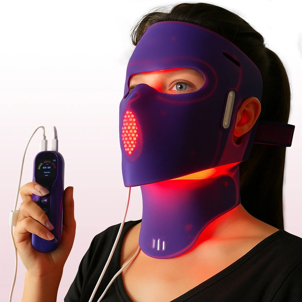
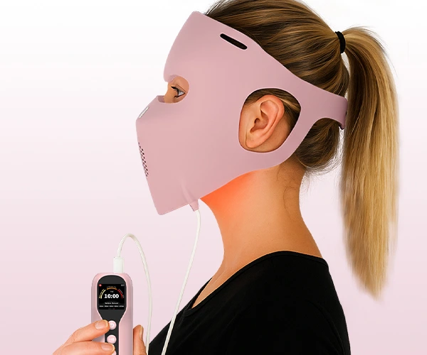
How WakeLife Beauty Implements Dual-Wavelength Design
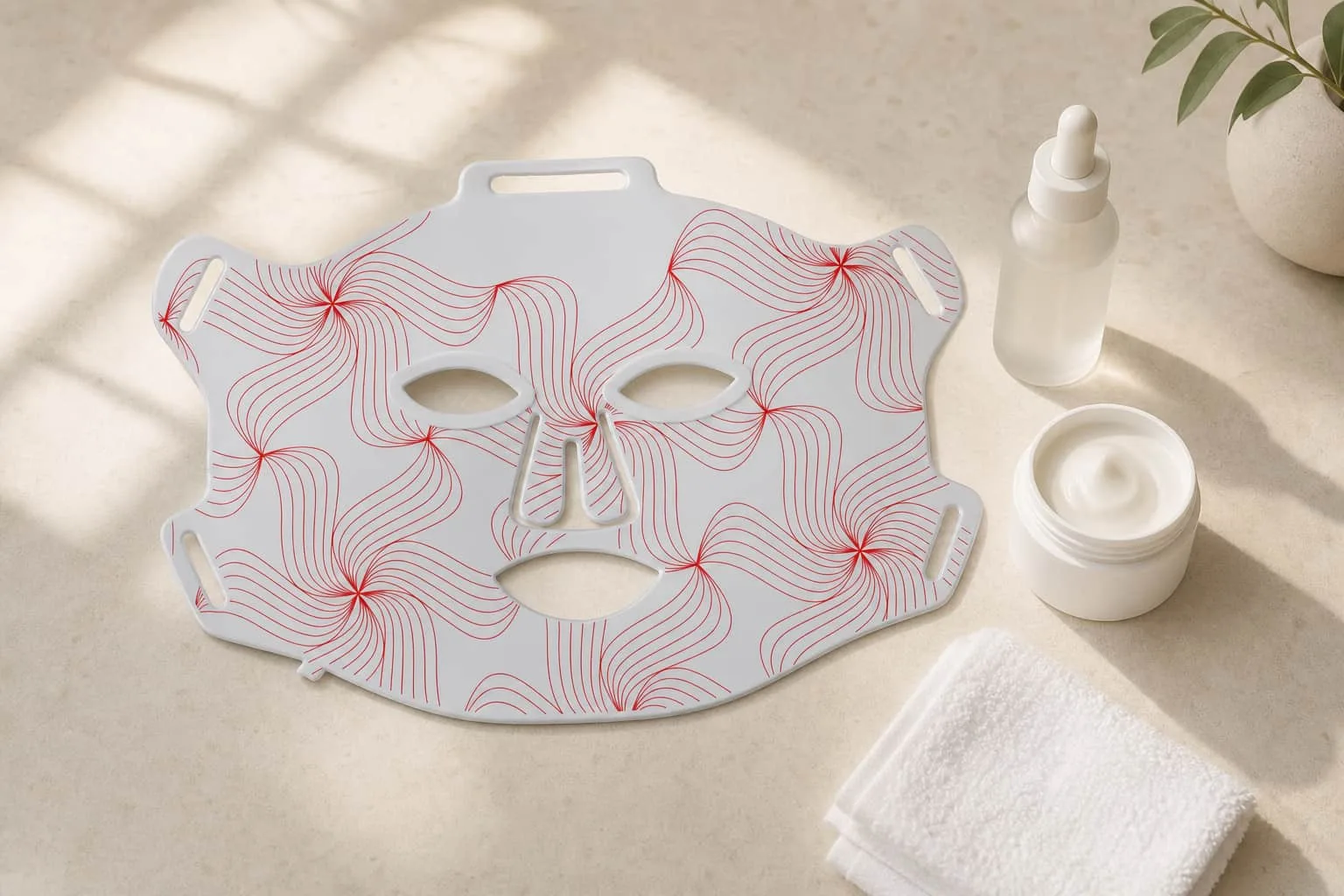
Our G15 LED Face Mask uses an optimized dual-wavelength configuration:
- 660 nm: Targets facial dermis for collagen stimulation and skin texture improvement
- 850 nm: Reaches deeper dermal layers and hair follicles for comprehensive rejuvenation
- Precision ratio: Red-to-NIR ratio calibrated for facial tissue anatomy (thin skin, high vascularity)
- Clinical-grade irradiance: Delivers therapeutic dose within practical treatment times
For OEM partners: WakeLife offers customizable wavelength configurations across our product line. Whether you need a skin-focused 660 nm device or a full-depth 660+850 nm panel, our engineering team can optimize the LED array for your target application.
Factors That Affect Penetration Depth Beyond Wavelength
Wavelength is the primary determinant, but several other factors influence how much light actually reaches the target:
Tissue-Side Factors
| Factor | Effect | Clinical Implication |
|---|---|---|
| Skin pigmentation | Higher melanin → more absorption at 400–700 nm | Darker skin types may benefit more from NIR (800+ nm) which bypasses melanin |
| Blood perfusion | More blood → more hemoglobin absorption | Highly vascular areas (face, scalp) absorb more red light |
| Tissue density | Dense tissue (muscle, bone) scatters more than fat | Fat is relatively transparent; muscle attenuates more |
| Hydration | Water absorbs above 970 nm | Primarily affects wavelengths >900 nm; minimal impact on 660/830 nm |
| Age | Aging changes collagen structure, skin thickness | Older skin may have slightly different optical properties (Bashkatov et al., 2005) |
Device-Side Factors
| Factor | Effect | Best Practice |
|---|---|---|
| Irradiance (mW/cm²) | Higher irradiance → more photons at depth (but does NOT change penetration depth itself) | 30–100 mW/cm² at tissue surface for most applications |
| Contact vs. non-contact | Direct contact eliminates surface reflection (4–7% loss) | Contact mode improves light coupling |
| Angle of incidence | Perpendicular = maximum transmission | Maintain 90° angle to tissue |
| Treatment distance | Follows inverse square law for non-contact devices | Consistent distance is critical for consistent dosing |
| Beam area | Larger area = more total energy delivered | Consider coverage area in protocol design |
Common misconception: Higher irradiance does NOT make light penetrate deeper. A 100 mW/cm² device and a 30 mW/cm² device at 660 nm penetrate to the same depth — the difference is that more photons arrive at that depth per unit time with higher irradiance, reaching therapeutic dose faster.
How to Choose: Wavelength Selection by Application
Decision Framework for Device Designers
| If your target is… | Primary wavelength | Consider adding | Rationale |
|---|---|---|---|
| Facial skin | 660 nm | 830/850 nm | Collagen in dermis + deeper stimulation |
| Acne | 660 nm (+ blue 415 nm) | 830 nm | Red for inflammation; blue for P. acnes bacteria |
| Wrinkles / fine lines | 660 nm | 830 nm | Multi-depth collagen remodeling (Wunsch & Matuschka, 2014) |
| Hair regrowth | 660 nm | 850 nm | Follicles sit at 2–5 mm depth |
| Wound healing | 660 nm | 830 nm | Surface repair + deep tissue regeneration |
| Muscle recovery | 830/850 nm | 660 nm | Deep penetration primary; red for surface circulation |
| Joint pain | 830/850 nm | 660 nm | Must penetrate through skin + fat to synovium |
| Bone healing | 830/850 nm | — | 10–30 mm penetration required |
| Brain / neurological | 810 nm | — | Optimal transcranial penetration; most research data |
Decision Framework for Consumers / Clinicians
Ask one question: How deep is my target tissue?
- Surface (0–5 mm) → Skin, wounds, acne, hair → Red (660 nm) is sufficient
- Deep (5–50 mm) → Muscle, joints, bone, nerve → NIR (830/850 nm) required
- Both → Full-body wellness, multi-condition → Dual-wavelength (660 + 830/850 nm)
If your condition is not improving with red light alone, the most common reason is insufficient penetration depth — switching to or adding NIR may solve the problem.
FAQ
What is the best wavelength for red light therapy?
It depends on your target. For skin conditions: 660 nm. For deep tissue (muscle, joints): 830 nm. For comprehensive treatment: dual-wavelength 660 + 830 nm.
How deep does 660 nm red light actually penetrate?
Approximately 1–5 mm effectively. This covers the full dermis and superficial hair follicles but is insufficient for muscle or joint targets.
Does skin color affect which wavelength to choose?
Yes. Darker skin contains more melanin, which absorbs visible red light (630–660 nm) more strongly. NIR wavelengths (800+ nm) are less affected by melanin and may be more effective for deeper treatment in darker skin types.
Why do some devices use 850 nm instead of 830 nm?
850 nm LEDs are more widely manufactured and cost-effective. The biological difference between 830 nm and 850 nm is minimal — both effectively target the CuA center of cytochrome c oxidase.
Can I combine red and NIR wavelengths?
Yes, and it is increasingly the clinical standard. Dual-wavelength devices activate both CcO absorption sites (heme center and CuA center) and treat across multiple tissue depths simultaneously.
Does higher power make light penetrate deeper?
No. Higher irradiance delivers more photons at the same depth, but does not change the physical penetration depth. A 100 mW/cm² device at 660 nm reaches the same depth as a 30 mW/cm² device at 660 nm — it just delivers the therapeutic dose faster.
Conclusion
Wavelength is not a marketing detail — it is the physics that determines whether your device can work for a given application.
The core principle is simple:
- 660 nm → Skin and superficial tissue (1–5 mm)
- 830/850 nm → Muscle, joints, and deep tissue (10–50 mm)
- 660 + 830 nm → Comprehensive coverage across all depths
For device manufacturers evaluating wavelength configurations for new products, WakeLife Beauty provides OEM/ODM services with customizable wavelength options, clinical-grade LED arrays, and engineering support for optimal therapeutic design.
Related Topics
References
Jacques, S.L. (2013). Optical properties of biological tissues: a review. Physics in Medicine & Biology, 58(11), R37-R61. PubMed
Karu, T.I., Pyatibrat, L.V., & Kalendo, G.S. (2004). Photobiological modulation of cell attachment via cytochrome c oxidase. Photochemical & Photobiological Sciences, 3(2), 211-216. PubMed
Wunsch, A., & Matuschka, K. (2014). A controlled trial to determine the efficacy of red and near-infrared light treatment in patient satisfaction, reduction of fine lines, wrinkles, skin roughness, and intradermal collagen density increase. Photomedicine and Laser Surgery, 32(2), 93-100. PubMed
Salehpour, F., et al. (2019). Penetration profiles of visible and near-infrared lasers and light-emitting diode light through the head tissues in animal and human species: a review of literature. Photobiomodulation, Photomedicine, and Laser Surgery, 37(10), 581-595. PubMed
Mochizuki-Oda, N., et al. (2002). Effects of near-infra-red laser irradiation on adenosine triphosphate and adenosine diphosphate contents of rat brain tissue. Neuroscience Letters, 323(3), 207-210. PubMed
Ferraresi, C., Huang, Y.Y., & Hamblin, M.R. (2016). Photobiomodulation in human muscle tissue: an advantage in sports performance? Journal of Biophotonics, 9(11-12), 1273-1299. PubMed
Bashkatov, A.N., Genina, E.A., Kochubey, V.I., & Tuchin, V.V. (2005). Optical properties of human skin, subcutaneous and mucous tissues in the wavelength range from 400 to 2000 nm. Journal of Physics D: Applied Physics, 38(15), 2543-2555. IOP Science